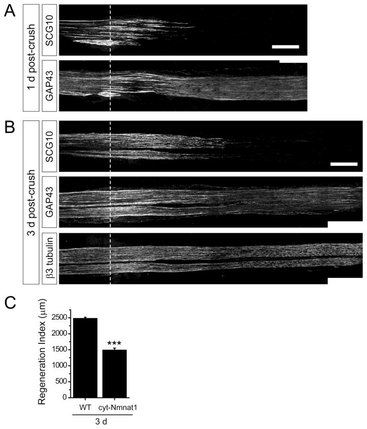
Figure 7. Selective SCG10 labeling reveals slowed axon regeneration in cyt-NMNAT1 mice.
(A) Distribution of SCG10 and GAP43 are compared at 1 d after crush in cyt-NMNAT1 mice, a model of delayed Wallerian degeneration. The distal axons are devoid of SCG10 despite of the slowed axon degeneration program. GAP43 robustly stains the distal axons. Scale bar = 500 μm.
(B) At 3 d after crush, the sciatic nerve of cyt-NMNAT1 is immunostained for SCG10, GAP43 and β3 tubulin. SCG10 staining shows that neurons from cyt-NMNAT1 expressing mice display slowed axon regeneration (quantified in (C)). GAP43 labeling persists in the distal axon segments. β3 tubulin shows that the distal cyt-NMNAT1 axons are intact at 3 d after injury. Scale bar = 500 μm.
(C) Axon regeneration in cyt-NMNAT1 mice are quantified by regeneration indices obtained from SCG10 immunostaining at 3 d after crush injury. n = 3 for each condition; ***p < 0.001 by t test. Error bars represent SEM.

