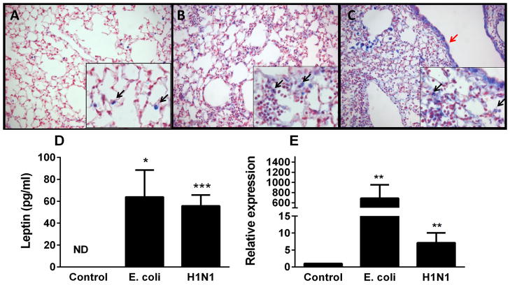
Figure 3.
Leptin levels are increased in murine lungs after bacterial or viral infection. Immunohistochemical examination was performed for leptin in normal murine lung tissue (n=5) (A), murine lung tissue infected with E. coli (n=5) (B), and murine H1N1 infected lung tissue (n=5) (C). Leptin is indicated by blue staining and nuclei are counterstained in red. Leptin staining was localized in alveolar macrophages (black arrows) and alveolar epithelium (red arrows). Original magnification x 200 (inserts x 400). Leptin levels were determined in either brochoalveolar lavage by ELISA (D) or lung tissue by qPCR (E) from mice exposed to either saline control (n=4), E. coli for 24h (n=8) or H1N1 for 4d (n=4). Data are presented as mean ± SEM, * p≤0.05, ** p≤0.01 and ***p≤0.001 compared to control.

