Figure 4.

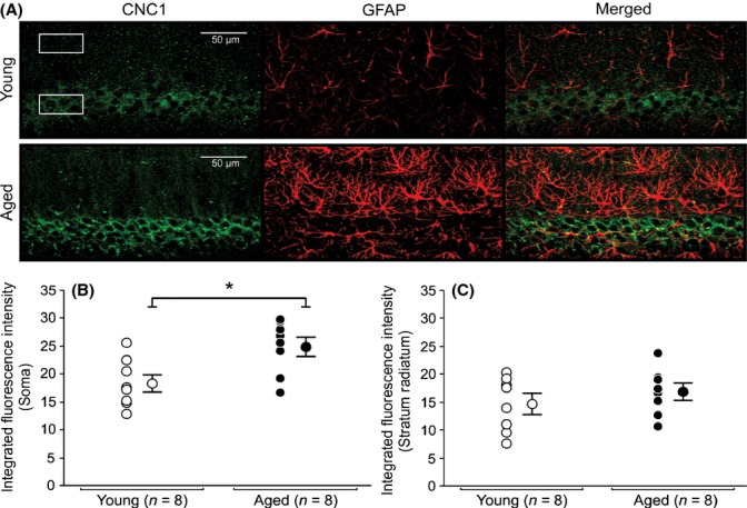
Expression of Cav1.2 L-type subunit in soma and radiatum of young and aged CA1 pyramidal neurons. (A) Representative confocal images of hippocampal CA1 pyramidal layer sections showing immunohistochemical labeling for Cav1.2 (CNC1) of a young and an aged rat. Six regions of interest (box) with equal dimensions in both the stratum pyramidale (3) and the stratum radiatum (3) layers of CA1 were drawn to collect immunofluorescence data. (B) Quantitative analysis of integrated fluorescence intensity in soma of CA1 pyramidal neurons of young and aged rats. (C) Quantitative analysis of integrated fluorescence intensity in the stratum radiatum of young and aged rats. Significant increases in somatic expression of Cav1.2 subunit were observed in aged CA1 pyramidal neurons (B) (P < 0.05). No significant differences in Cav1.2 subunit expression were detected between stratum radiatum of young and aged rats. No colocalization of CNC1 was observed in glial cells. AutoQuant image deconvolution software (Media Cybernetics, Rockville, MD) was used to reduce background signal for the purpose of illustration. Fluorescence intensities and analyses were performed using raw, unmodified, images. Data reported as the mean ± SEM.
