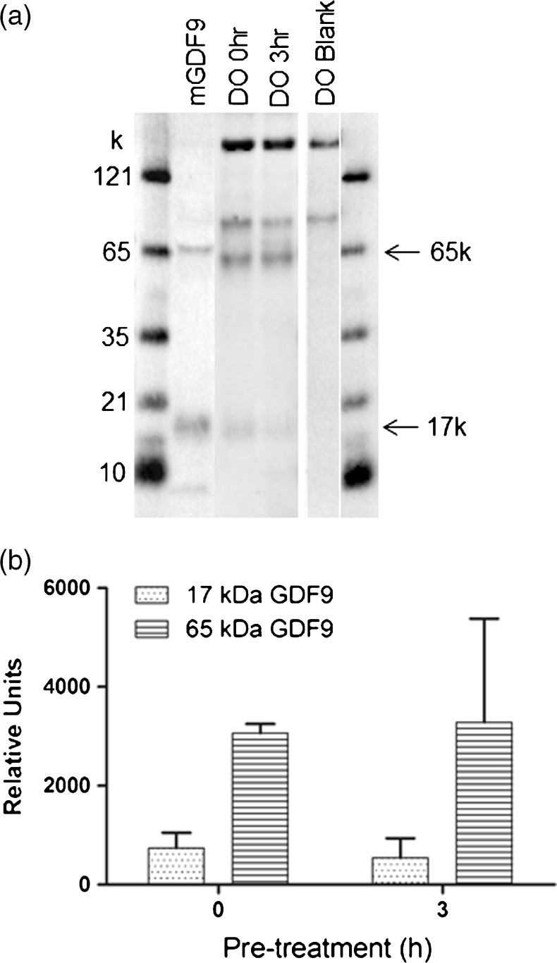
Fig. 2.
a GDF9 Western blot comparison of denuded oocyte (DO) extracts at 0h and 3 h (3 h + FSH + CC) of pretreatment. mGDF9 refers to recombinant mGDF9 standard. Blank refers to DO sample in the absence of the GDF9 primary antibody. The 65 kDa band corresponds to the unprocessed GDF9 pro-protein while the 17 kDa band corresponds to the mature domain. The 90 kDa–180 kDa bands seen in all samples are attributed to the non-specific presence of biotinylated proteins in the cell extracts b Quantitative analysis of Western blot denuded oocyte extracts at 0 and 3h (3 h + FSH + CC) of pretreatment from 5 separate experiments. The Western blot image was quantified using BioRad ChemiDoc MP system and analysed using Image J software (Biorad)

