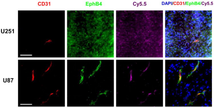
Fig. 7.
Fluorescence microscopy of mouse brains with implanted U87-Luc or U251-Luc tumors excised after imaging studies. Mice bearing a U251-Luc or U87-Luc tumor were injected intravenously with Cy5.5-TNYL-RAWK-64Cu-DOTA peptides. Signal from Cy5.5 is pseudocolored purple. EphB4 (green) was stained with rabbit anti-human EphB4 antibody and Alexa Fluor 488-conjugated goat anti-rabbit immunoglobulin. CD31 (red), which is the marker of the endothelial cells of tumor-associated blood vessels, was stained with rat anti-mouse CD31 antibody and Alexa Fluor 594-conjugated donkey anti-rat antibody. Cell nuclei were counterstained with DAPI (blue). Bar=40 μm.

