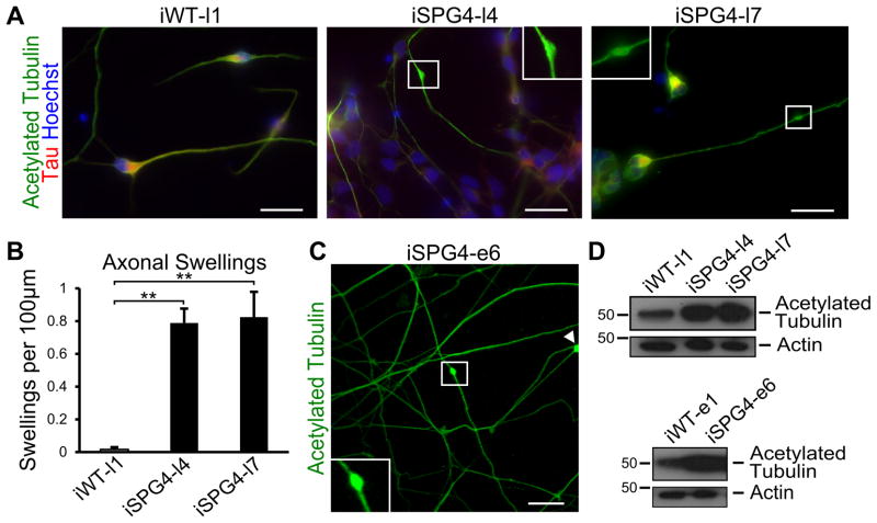
Figure 2.
SPG4 patient-derived neurons display axonal defects. (A) Acetylated tubulin staining revealed the presence of swellings along Tau+ axons of 6 week-old neurons. Boxed areas are enlarged in insets. (B) Quantification revealed a significant increase in axonal swellings in patient-derived neurons compared to control neurons. Data presented as mean ±SD. **P < 0.01. (C) Episomal SPG4 neurons also possessed axonal swellings. (D) Western blots show that acetylated tubulin levels were dramatically increased in the week 6 SPG4-derived neurons compared to controls. Scale bars: 20 μm.

