Figure 4.
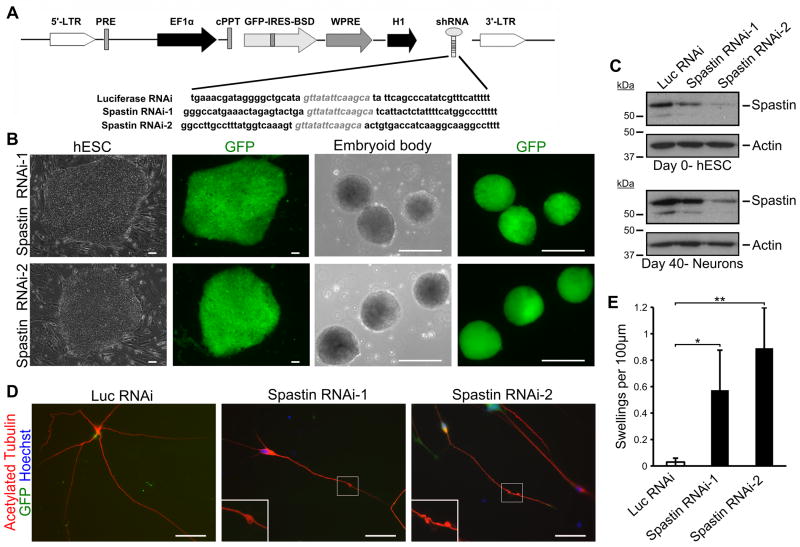
Spastin knockdown in hESC-derived neurons recapitulates SPG4 patient phenotype. (A) Schematic map showing the pLVTHM vector with the shRNA sequence. H9 hESCs were transfected with the indicated lentivirus, and GFP+ cells were selected. (B) hESC colony after lentiviral transfection shows efficient generation of knockdown lines. Expression of shRNA continued during differentiation. Scale bars: 100 μm. (C) Western blot analysis confirming knockdown of spastin in both hESCs and neurons after 40 days of differentiation. (D) Immunostaining for acetylated tubulin revealed the development of axonal swellings in two spastin RNAi hESC-derived neurons. Hoechst stains the nuclei. Scale bars: 20μm. (E) Quantification of axonal swellings shows a significant increase in spastin RNAi-derived neurons compared to control cells. Data presented as mean ±SD. *P < 0.05, **P < 0.01. Luc, luciferase.

