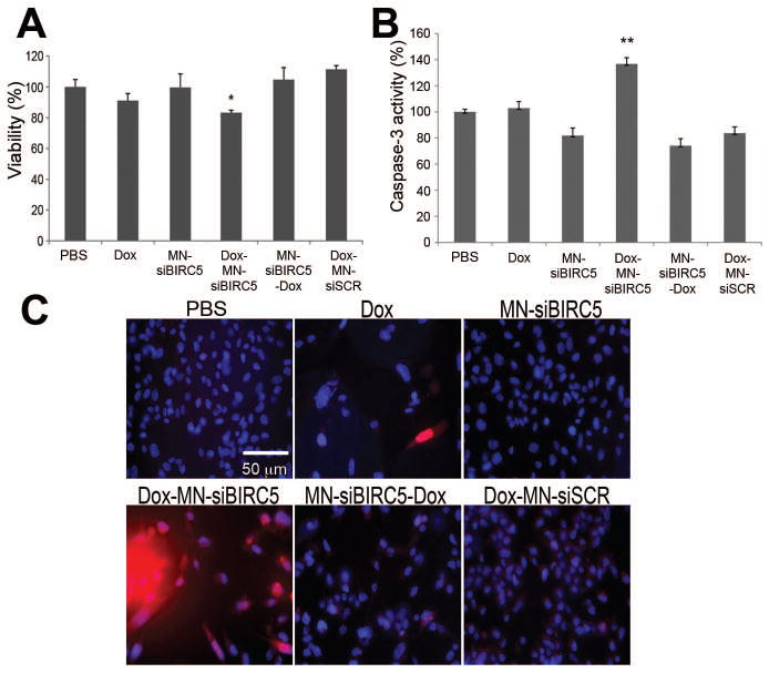
Fig. 1. In vitro pro-apoptotic effects of Dox and MN-siBIRC5 in breast adenocarcinoma (BT-20) cells.
(A) Effect of the drugs on cell proliferation, as evaluated by MTT assay. BT-20 cells were treated with PBS, Dox (0.1μM) or nanodrug either alone or in sequential combination (Dox-MN-siBIRC5: Dox alone for 24h and then MN-siBIRC5 for another 24 h; MN-siBIRC5-Dox: MN-siBIRC5 for 24h and then Dox for another 24 h). Results are expressed as mean ± SD and represent a summary of three independent experiments (n=3, two-tailed Student’s t-test, *P<0.001). (B) Effect of the drugs on caspase-3 activation. BT-20 cells were treated with PBS, Dox (0.1μM) or nanodrug either alone or in sequential combination (Dox-MN-siBIRC5: Dox alone for 24h and then MN-siBIRC5 for another 24 h; MN-siBIRC5-Dox: MN-siBIRC5 for 24h and then Dox for another 24 h). Caspase-3 activity was measured in cell lysates prepared 48h after additional incubation with the drugs. Caspase-3 activation was significantly higher in the Dox-MN-siBIRC5 treatment group relative to PBS. Data are expressed as mean ± SD and are representative of three independent experiments (n=3; two-tailed Student’s t-test, *P<0.05, **P<0.005). (C) Analysis of PARP cleavage. After exposing the cells to drug treatments (as above), immunofluorescence was performed to detect cleaved PARP fragments. Nuclei (DAPI, Blue) and cleaved PARP (Red) were visualized by fluorescence microscopy. PARP cleavage was most prominent in the Dox-MN-siBIRC5 treatment group.

