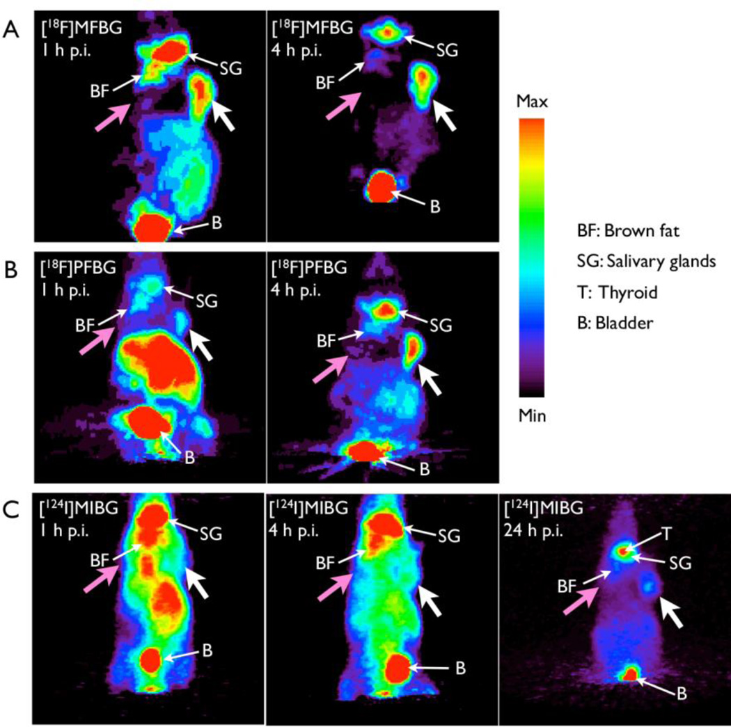
Figure 4.
Typical PET projection images of [18F]MFBG, [18F]PFBG and [124I]MIBG in animals bearing dual xenografts (C6-hNET (right) and C6-WT (left)). [18F]MFBG (A) and [18F]PFBG (B) PET imaged at 1 h and 4 h p.i., respectively; (C) PET images of [124I]MIBG at 1 h, 4 h and 24 h post injection. The projection images are from the same animals shown in Figure 3. A color threshold was optimized to visualize the C6-hNET tumor (white arrow) on the projection image; the C6 wild-type tumor (pink arrow) was not visualized.

