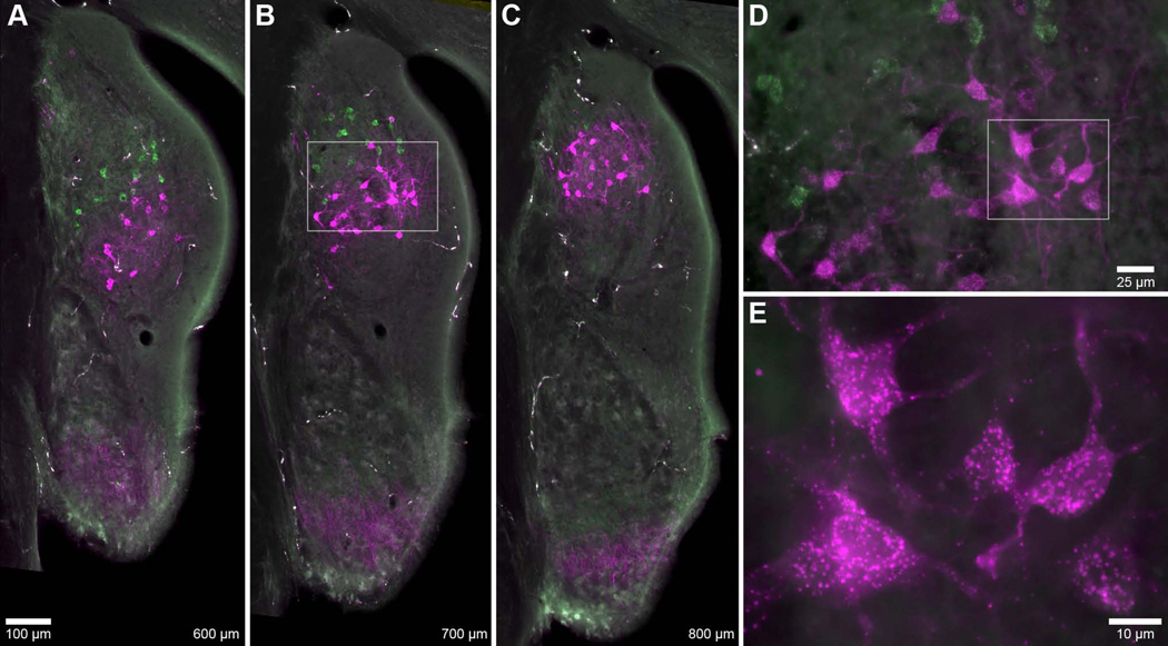
Figure 3.
Photomicrographs of labeled vertical cells. A–C: Coronal sections through the DCN and PVCN spaced 100 µm apart. AN fibers and vertical cells were labeled by the pair of injections shown in Figure 1 (#38, Table 1). Vertical cells (and faint AN fibers) matched to the higher BF injection (green, 17.7 kHz; magenta, 10.0 kHz) are consistently located more posterodorsally. Cells are distributed throughout the deep layers of the DCN, and do not occupy superficial layers. Values at bottom-right designate relative distance from the posterior pole of the CN. Scale bar in A applies to A–C. D: High magnification of vertical cells from box in B. The longer branches of vertical cell dendrites tended to extend towards the superficial layers (upper-right). E: High magnification of vertical cells from box in D. Cell bodies were slightly elongated and variable in shape, consistent with previous descriptions of vertical cells (Zhang and Oertel, 1993; Rhode, 1999).

