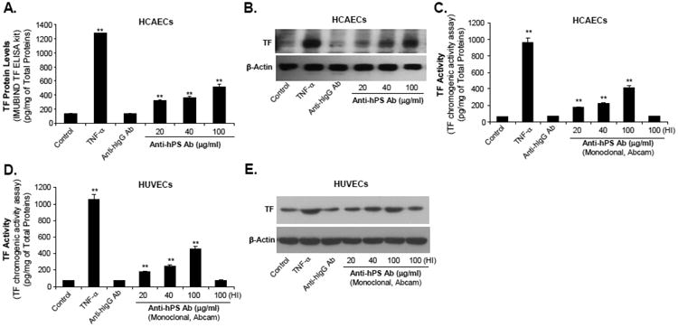
Fig. 2.

Effect of anti-hPS Ab on TF protein levels as well as TF-mediated procoagulant activity in HCAECs and HUVECs. (A). Concentration-dependent response of anti-hPS Ab-induced TF protein levels (ELISA) in HCAECs. Serum-starved HCAECs were treated with 20-100 μg/mL anti-hPS Ab (polyclonal, Sigma), 100 μg/mL anti-hIgG Ab or 5 ng/mL TNF-a for 3 hours. The cellular TF protein levels were measured by IMUBIND Tissue Factor ELISA Kit. (B). Concentration dependent response of anti-hPS Ab-induced TF protein levels (Western blot) in HCAECs. Serum-starved HCAECs were treated with 20-100 μg/mL anti-hPS Ab, 100 μg/mL anti-hIgG Ab or 5 ng/mL TNF-α for 3 hours. TF protein expression was detected by Western blot. β-actin was used as a loading control. (C). Effect of anti-hPS Ab (monoclonal, Abcam) on TF-mediated procoagulant activity in HCAECs. Serum-starved HCAECs were treated with 20-100 μg/mL anti-hPS Ab, 40 μg/mL anti-hIgG Ab, 5 ng/mL TNF-a or heat-inactivated anti-hPS Ab (100 ng/mL) for 3 hours. The TF activity levels were measured by a tissue factor human chromogenic activity assay kit (Abcam). (D). Effect of anti-hPS Ab (monoclonal, Abcam) on TF-mediated procoagulant activity in HUVECs. (E). Effect of anti-hPS Ab (monoclonal, Abcam) on TF expression (Western blot) in HUVECs. Data shown are the mean + SEM of triplicate determinations. **P < 0.01 versus untreated controls.
