Fig. 3.

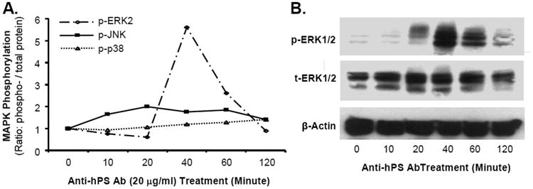
Effect of anti-hPS Ab on MAPK phosphorylation in HCAECs. (A). The activation status of MAPKs (ERK2, JNK and p38) was analyzed by Bio-Plex immunoassay. Serum-starved HCAECs were treated with anti-hPS Ab (20 μg/mL) and the cell lysates were harvested at different time points with Bio-Plex Cell Lysis Kit. The phosphoprotein and total proteins of MAPKs were analyzed by a Luminex 100TM analyzer and Bio-Plex Manager software (BioRad). (B). Phosphorylation of ERK1/2 was detected by Western blot. Serum-starved HCAECs were treated with anti-PS Ab (20 μg/mL) for different time points. The phosphorylated and total ERK1/2 proteins were detected by Western blot. β-actin was used as a loading control. Representative results from 3 experiments are shown.
