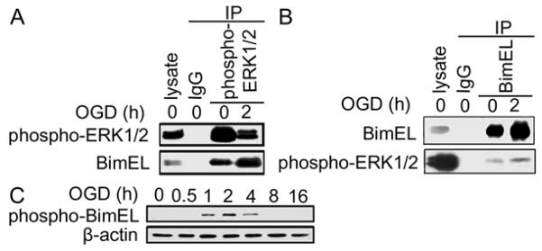
Figure 5.

Interaction of BimEL with phospho-ERK1/2. (A) Co-immunoprecipitation of BimEL with phospho-ERK1/2. SH-SY5Y cells were exposed to OGD and then lysed and extracts were collected. The extracts were subjected to immunoprecipitation with IgG (control) or anti phospho-ERK1/2 and followed by Western blot analyses with anti-phospho-ERK1/2 or anti-BimEL antibody. (B) Reverse immunoprecipitation with BimEL and detected for phospho-ERK1/2. SH-SY5Y cells were exposed to OGD and then lysed and extracts were collected. The extracts were subjected to immunoprecipitation with IgG (control) or anti-BimEL and followed by Western blot analyses with anti-phospho-ERK1/2 or anti-BimEL antibody. (C) phospho-BimEL expression during OGD. Cells were treated with OGD for the indicated hours. Endogenous level of phospho-BimEL was detected with Western blot analysis. The experiments were repeated two to three times with similar results.
