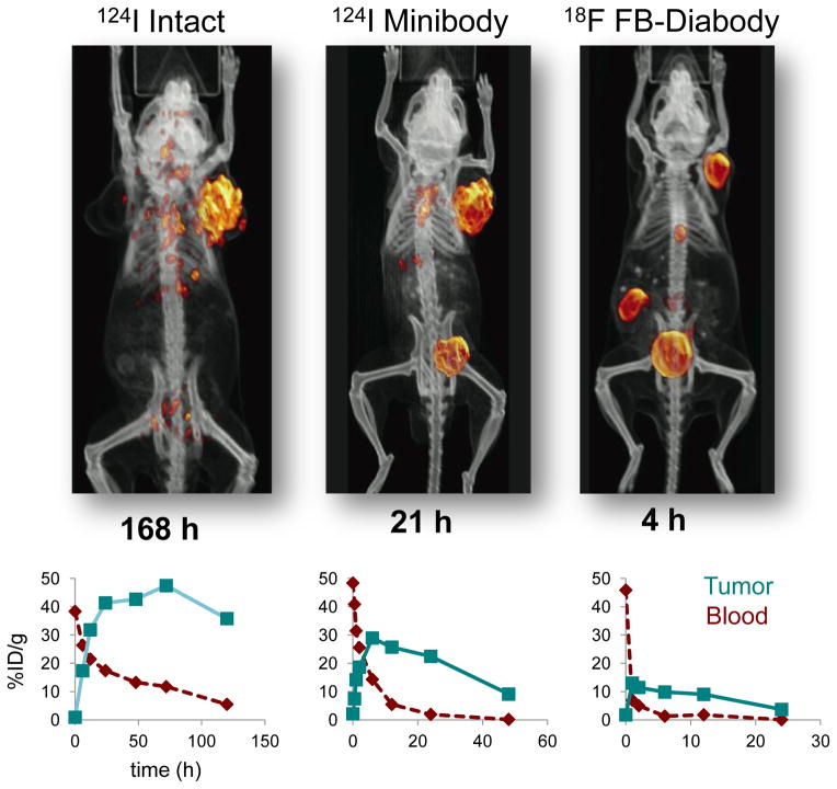
Figure 3.
Accelerated targeting, clearance, and imaging afforded by minibody and diabody formats compared to intact antibodies. Top row shows representative microPET images using 124I- radiolabeled intact antibody and minibody, and 18F-radiolabeled diabody recognizing prostate stem cell antigen (PSCA) in athymic mice bearing LAPC-9 human prostate cancer tumor xenografts [2, 26, 27]. Imaging times post injection are indicated below each image. Bottom row shows typical tumor and blood biodistribution curves for radiolabeled intact, minibody, and diabody formats in s.c. xenograft models in mice (anti-CEA in LS174T human colon cancer tumors) [21].

