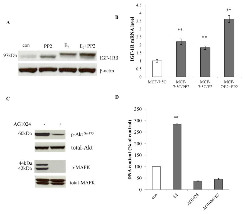
Figure 5. The c-Src inhibitor collaborated with E2 to elevate IGF-1Rβ.
(A) IGF-1Rβ changes after long-term treatment. Cell lysates of cells treated in different combinations were harvested. IGF-1Rβ was examined by immunoblotting. β-actin was detected for loading control. (B) IGF-1Rβ mRNA changes were consistent with protein levels. The RNA of differently long-term treated cells was harvested as above. P<0.001, ** compared with control. (C) Activation of Akt and MAPK pathways by IGF-1Rβ in MCF-7:PF cells. MCF-7:PF cells were treated with vehicle (0.1% DMSO) and AG1024 (10−5mol/L) for 48 hours. Cell lysates were harvested. Phosphorylated Akt and MAPK were determined by immunoblotting. Total Akt and MAPK were examined for loading controls. (D) The IGF-1R inhibitor completely blocked E2 stimulation in MCF-7:PF cells. MCF-7:PF cells were treated with vehicle (0.1% EtOH), E2 (10−9mol/L), AG1024 (10−5mol/L), and E2 (10−9mol/L) plus AG1024 (10−5mol/L) for 7 days. The cells were harvested and DNA content was determined as above. P<0.001, ** compared with control. All the data shown were representative of at least three separate experiments with similar results.

