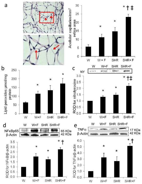
Fig. 1.
HFD causes retinal microvascular degeneration and oxidative and inflammatory stress. (a) Representative images and quantification of retinal trypsin digests with enlarged subset identifying retinal acellular capillaries (arrows) in control rats (W) and SHR fed with normal diet or with HFD (W+F) and (SHR+F), respectively, for 10 weeks. Magnification, ×200; scale bar, 25 μm. Two-way ANOVA showed significant interaction between HFD and hypertension. Oxidative stress markers were assessed using retinal lipid peroxidation levels (b) and NY levels (c) in all groups. Two-way ANOVA showed significant interaction between HFD and hypertension in retinal NY levels. Representative blots and western blot analyses of NFκB p65 expression (d) and TNF-α expression (e) in all groups. Protein levels were normalised to β-actin and respective control group. n=4 or 5; *p<0.05 vs W; †p<0.05 vs SHR; ‡p<0.05 vs W+F

