
Figure 1. Purity of nDNA and mtDNA.
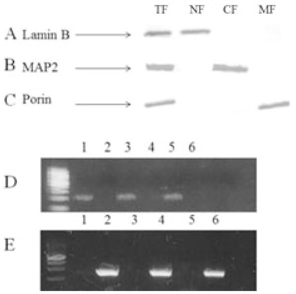
Representative Western blot analysis of total-(TF; Row 1), nuclear- (NF; Row 2), cytosolic- (CF; Row 3), and mitochondrial-fractions (NF; Row 4) probed for Lamin B a nuclear envelope protein (A), MAP2 a microtubular associated protein (B), and Porin a voltage dependent anion channel found on the outer membrane of the mitochondria (C). Representative PCR amplification product of APOE (D) and MT-ND2 (E) of mtDNA (Lane 1) and nDNA (Lane 2) from SMTG, mtDNA (Lane 3) and nDNA (Lane 4) from IPL, mtDNA (Lane 5) and nDNA (Lane 6) from CER.