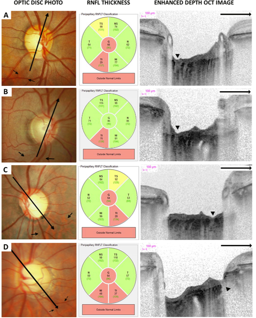
Figure 4.
Optic disc photographs, retinal nerve fiber layer (RNFL) thickness classification and multiple enhanced depth optical coherence tomography (EDI-OCT) radial scans of eyes with lamina cribrosa defects (arrow heads) corresponding to areas of localized RNFL loss (small arrows). The EDI-OCT radial scan direction is indicated (large arrows).

