Fig. 6.
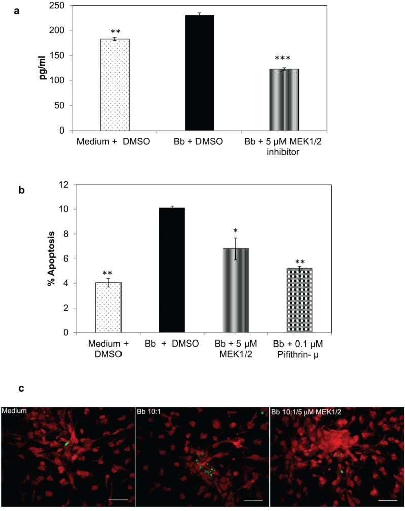
Effect of MEK pathway inhibition on inflammation and apoptosis of primary oligodendrocytes exposed to B. burgdorferi (MOI 10:1). 6a) B. burgdorferi-mediated IL-8 release from primary oligodendrocytes is suppressed by the inhibition of MEK pathway. A representative experiment is shown with bars representing standard deviation; ** p < 0.01;*** p < 0.001. 6b) and (6c) show the results of apoptosis as evaluated by TUNEL assay in primary human oligodendrocytes in response to B. burgdorferi ± inhibitors. After 48 h of exposure to B. burgdorferi, cells were stained with MBP (red) and processed for TUNEL as described in Materials and Methods. Nearly 2000 (or more) cells were counted/well/treatment over 13-20 microscope fields along with TUNEL positive cells (green) in each field. 6b) shows the mean apoptotic values from 2 independent but identical experiments with bars representing SEM. * p < 0.05; ** p < 0.01. Panels in (6c) show apoptosis (green) in differentiated HOPC cells (red) for various treatment groups. Bar represents 100 μm

