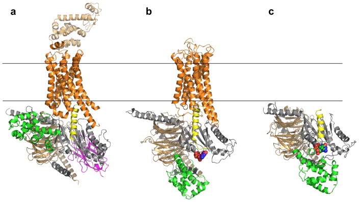
Figure 1.

Overall structure of β2AR–Gs complex, our model of the R*–Gi complex, and the unbound Gi heterotrimer. (a) Crystal structure of β2AR–Gs complex (PDB 3SN611). The α5 helix of Gas is displaced 6Å towards the receptor and the helical domain (green) is displaced towards the membrane interface. (b) Unified model of the R*–Gi complex: According to DEER measurements, the displacement of helical domain (green) is on average a 15Å translation and 62º rotation after receptor binding. (c) Gi heterotrimer constructed as comparative model from Gt (PDB 1GOT10) structure. Receptor (orange), Gα GTPase domain (grey), Gα helical domain (green), Gβ (light brown), Gγ (black), Nanobody (magenta), T4L (sand), GDP (in spheres).
