
Figure 3. IL-6 drives experimentally induced M. avium-IRIS.
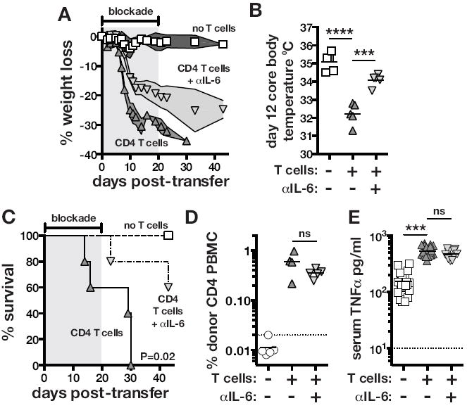
TCRαKO mice were intravenously injected with 1×10⁶ CFU of M. avium. After at least 2 months post-infection, 2×10⁶ WT CD4 T cells were adoptively transferred intravenously, and where indicated mice were then either untreated or administered IL-6 neutralizing mAb. (A) Weight loss was normalized to the day of T cell transfer. The shaded region indicates the duration of IL-6 blockade. (B) Core body temperature was measured on day 12 after T cell transfer. Uninfected TCRαKO mice displayed a body temp of 37-37.5°C. (C) Mortality was monitored following CD4 T cell reconstitution. The shaded region indicates the duration of IL-6 blockade. Data are representative of six independent experiments. (D) The frequency of donor CD4 T cells in the blood was measured on day 10 post-transfer. The data are representative of two independent experiments. (E) TNF serum concentrations were measured on day 11-12 post-transfer of CD4 T cells. The data are pooled from 4 independent experiments.