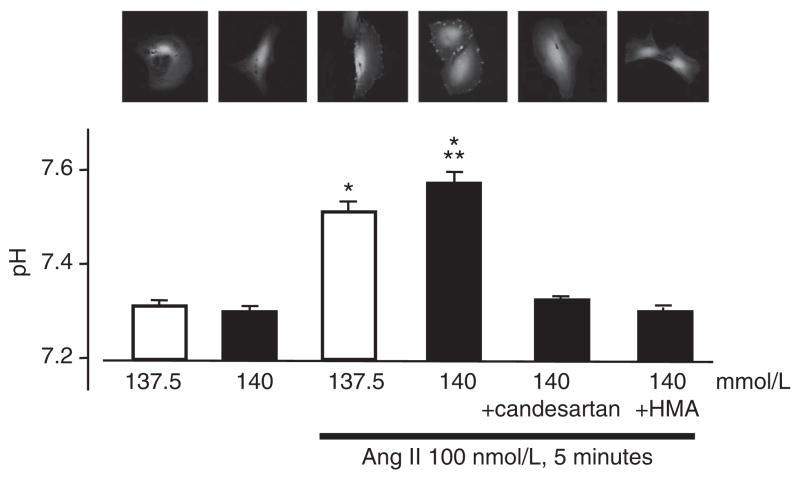
Figure 5.
Effect of sodium on intracellular pH in angiotensin II (Ang II)-treated vascular smooth muscle cells (VSMCs). Cells were incubated with the pH-reporter dye carboxy-SNARF-1. The pHi dye was excited with a 488 nm argon laser, and fluorescence was detected by confocal microscopy at 580 and 640 nm. Intracellular pH was evaluated by the fluorescence ratio using a standard curve. Data represent the mean±s.e. (n = 4). *P<0.05 vs. control VSMCs. **P<0.05 vs. Ang II-treated VSMCs in normal sodium medium. A full color version of this figure is available at the Hypertension Research journal online.

