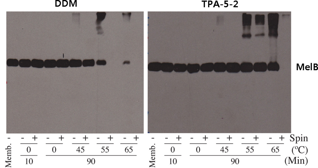
Figure 3.
Western blot analysis of MelB. Samples (10 µg) were separated by SDS-12% PAGE, and MelB was detected using an anti-histidine tag antibody. The solubilization extracts in TPA-5-2 and DDM were divided into two samples; one after ultracentrifugation (+) and one prior to ultracentrifugation (−). As a control, an untreated membrane sample (“Memb”; no ultracentrifugation) was included in each gel.

