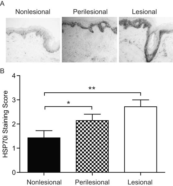
Figure 1.
HSP70i overexpression in vitiligo skin. (A) Representative immunoperoxidase staining of HSP70i in vitiligo skin displaying expression predominantly located in the epidermis, with minimal cellular expression observed in nonlesional (left), and moderate to strong expression in perilesional (middle) and lesional (right) skin. (B) Subjective, blinded quantification of HSP70i expression in tissue sections. Immunoperoxidase intensity: 1=low, 2=medium, 3=high. Data are presented as mean ± SEM as calculated by Student's t-test. *P< 0.05, **P<0.01, n=7.

