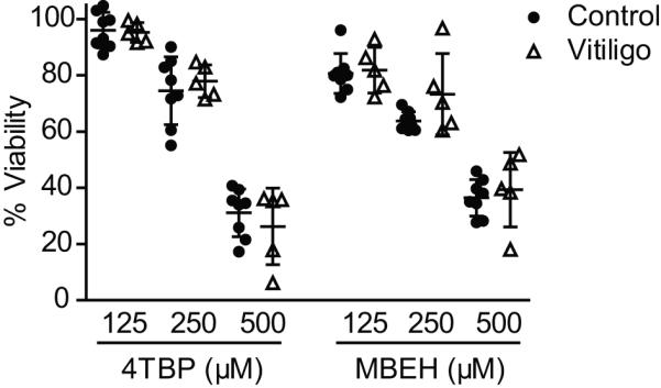
Figure 2.

Cell viability of vitiliginous melanocytes. Primary melanocytes from control and vitiligo patients were plated at 10,000 cells per well in triplicate, and exposed to 125, 250, or 500 μM concentration of 4-tertiary butyl phenol (4-TBP) or monobenzyl ether of hydroquinone (MBEH) for 72 hours. Percentage viability was quantified in MTT assays, and compared relative to vehicle treated controls. Cell viability decreased as 4-TBP and MBEH dosage increased; however, no differences were observed between control and vitiligo melanocytes for any treatment. Data provided from two independent experiments with triplicate values for each experiment. Healthy control (n =8), vitiligo (n = 5). Data are presented as mean ± SEM as calculated by Student's t-test.
