Figure 3.
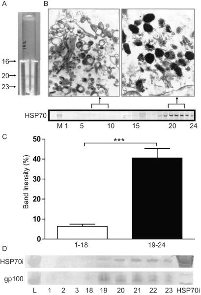
HSP70i colocalizes with melanosomal fractions. (A) Adult melanocytes (HM162P7) were dounced and underloaded on an iodixanol gradient for ultracentrifugation. Dense bands containing melanin were observed in the melanosomal (20/21 and 23) fractions. (B, upper image) Collected fractions (non-melanosomal 7–9, and melanosomal 20–22) were fixed and analyzed by electron microscopy (EM). (B, lower image) Collected fractions were run on a Western Blot. SPA-820 antibody (which binds both constitutive and inducible HSP70 isoforms) reacted with the melanosomal (19–24) but not nonmelanosomal fractions (1–15). (C) Quantification of western blot band intensities shown in EM. Mean luminosities indicate stronger antibody reactivity in fractions 19–24 versus 1–18. (D) Western blot analysis of non-melanosomal (1, 2 and 18) and melanosomal (19–23) fractions probed with the antibody SPA-810 which only detects inducible HSP70 (70 kD), and HMB45 to the melanocyte antigen gp100 (HMB45 detects a 45 kD product of gp100).Only the melanosomal fractions (19–23) react with both HSP70i and gp100 antibodies. Purified HSP70i protein was probed as a control. Together these data indicate that HSP70i is localized within the melanosome containing fractions.

