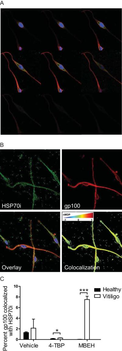
Figure 4.

HSP70i is overexpressed in vitiliginous melanosomes after stress. (A) Representative serial 1.0 μM Z-sliced images of neonatal melanocytes (Mf0627P11) indicate cytoplasmic expression of HSP70i (SPA-811 Ab detected by FITC) and the melanocyte antigen TRP-1 (Ta99 Ab detected by PE/Cy7) throughout the cell, but not present in the nucleus (DAPI counterstained). (B) Representative 0.5 μM Z-slice images of neonatal melanocytes (Mf0627P11) probed with antibodies to HSP70i (SPA-811) and gp100 (HMB45). Individual channels for HSP70i (GFP), gp100 (PE/Cy7) and merged images are shown. Note extracellular detection of HSP70i/gp100. An image pseudocolored by the ImageJ plug-in JACoP indicates levels of HSP70i/gp100 colocalization as low (blue), middle (yellow) and high (red). Perinuclear red mapping indicates over-lapping between the two labels, suggestive of colocalization. (C) Vitiligo and neonatal melanocytes were treated with 250 μM bleaching agents (4-TBP and MBEH) for 24 hours followed by confocal microscopy. Five z-slices from representative treated melanocytes were analyzed for HSP70i/gp100 colocalization using JACoP, with an nMDP cutoff of 1 used in the calculations. Data is presented as HSP70i/gp100 colocalization relative to total HSP70i staining. Graphed is the normalized mean deviation production (nMDP) for 5, 1μm Z-slices for each sample. Healthy control (n =3), vitiligo (n = 3). Data are presented as mean ± SEM as calculated by Student's t-test.* P<0.05, ***P< 0.001
