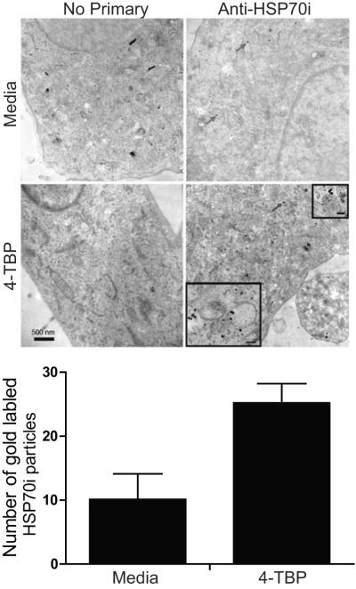
Figure 5.
Transmission electron microscope detection of HSP70i in 4-TBP treated melanocytes. Healthy melanocytes (Mf0632P2) were treated with 125 μM 4-TBP for 4 hours followed by immunostaining with gold particle-conjugated antibodies to HSP70i. The melanocytes were next fixed and scanned by transmission electron microscopy. Gold particles (red arrows) are observed in sections probed with anti-HSP70i which are more abundant (P < 0.0002) in 4-TBP treated melanocytes as shown in the bar graph below. HSP70i is seen throughout the cytoplasm in line with data shown in Figure 4, and occasionally juxtapositioned to melanosomes (insets). Bar equals 500 nm. (inset bars equal 40nm).

