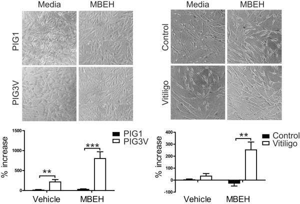
Figure 6.
Vitiligo melanocytes secrete more HSP70i in response to MBEH. (A) The immortalized melanocyte lines (vitiligo PIG3VP54 and control PIG1P96; n=3 measurements) and (B) primary healthy and vitiligo melanocytes were treated with 125 um MBEH for 24 hours. Supernatants from treated and untreated cultures were assessed for HSP70i content by high-sensitivity ELISA. The percent increase in HSP70i of MBEH and vehicle treated compared to untreated samples is indicated. Images of cell cultures (A and B) show no differences in cell density or death after MBEH treatment. A significant increase in HSP70i secretion was observed in vitiligo, but not control melanocytes after MBEH treatment. These data indicate that melanocytes obtained from vitiligo skin secrete more HSP70i in response to stress than healthy cells. Control (n =4 individual cultures), vitiligo (n = 3 individual cultures). Data are presented as mean ± SEM as calculated by Student's t-test. **P< 0.01, ***P< 0.001.

