
Figure 2.
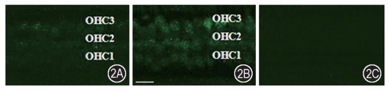
Nitrotyrosine increase in OHCs following noise exposure. A: Immunostaining of anti-nitrotyrosine in the organ of Corti of the control animals showed a relatively weak staining of NT in the outer hair cells. B: Significant NT immunostaining increase was observed in the outer hair cells of guinea pig cochleae following noise exposure. C: No immunoreactive staining was observed in control animals without the primary antibody. Scale bar = 30 μm.