
Figure 3.
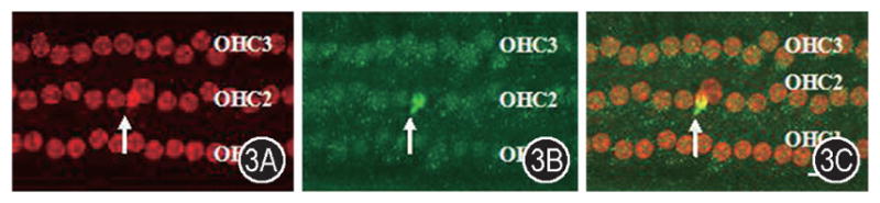
Anti-nitrotyrosine and propidium iodide double labeling showed the nitrotyrosine change in the outer hair cells after noise exposure. A: The organ of corti was stained with propidium iodide showing nucleoli after noise exposure. Apoptotic (arrow) OHCs were observed following noise exposure. B: Anti-NT antibody labeling shows the change of nitrotyrosine following noise exposure. A significant NT increase was observed in an OHC. C: Anti-NT antibody and propidium iodide double labeling shows that the apoptotic OHC with a condensed nuclei has a significant increase of nitrotyrosine in and around nucleus. OHC1, 2 and 3: first, second and third rows of outer hair cells. Scale bar = 20 μm.