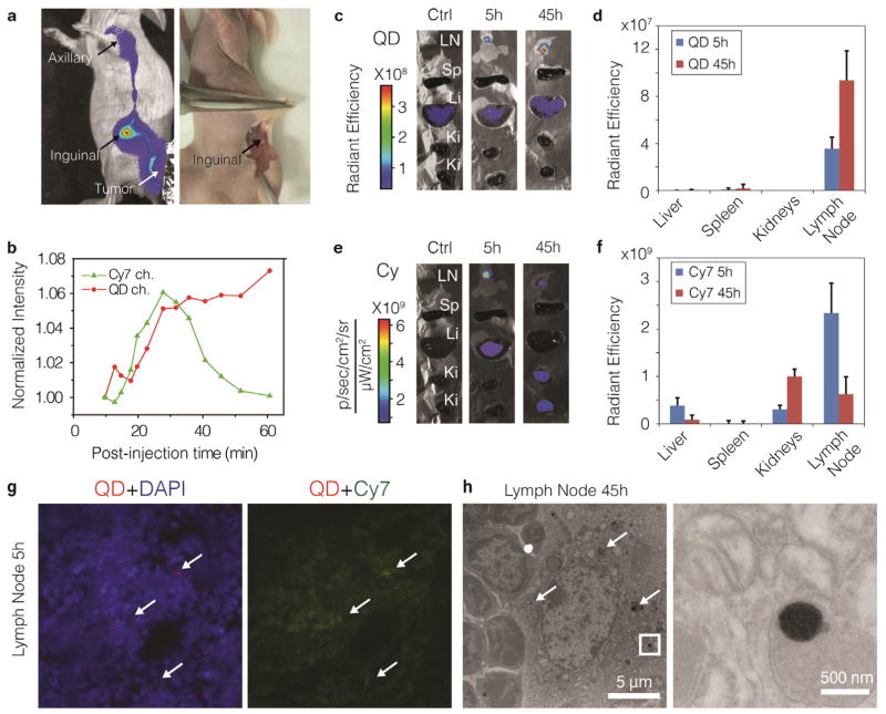
Figure 4. Peri-tumoral administration of lipid-coated nanocrystals.
a, NIR fluorescence image (left, laid over bright field image) show in both the QD and the Cy7 channel (Cy7 channel shown here), that the QD710-Cy7-PEG migrated through lymphatic draining from the periphery of the tumor to the inguinal node (as sentinel lymph node (SLN)) and further to the axillary node. After re-injection with 1% Evans blue, QD710-Cy7-PEG and Evans blue were found co-localized in the same lymph node, as indicated by the arrows (a, right, color image). b, Normalized total fluorescent intensities of QD710 (squares) and Cy7 (triangles) from the SLN are plotted against post-injection time, showing their different dynamic behaviours. Mice were sacrificed at 5 h and 45 h post-injection time (n=3 for each time point). Subsequently, the inguinal node and major organs were subject to ex vivo fluorescence imaging. Representative images and mean intensities are depicted in c and d for the QD channel and in e and f for the Cy7 channel. LN, lymph node. g, Fluorescence microscopy images of SLN tissue at 5 h post-injection. Merged images are shown with signal from QD (red), Cy7 (green) and DAPI (blue). Spots of QD accumulates are indicated with arrows. h, TEM images of SLN tissues at 45h after injection. Aggregates of QD cores inside the phagocyte are indicated by arrows. Insets are enhanced on the right.

