
Figure 5.
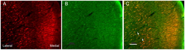
Microphotographs showing distribution and co-localization of GluN1 and μ opioid receptors in rACC neurons. A, B: Sections were double-labeled with guinea pig polyclonal antibody against μ opioid receptors (A, red) and goat polyclonal antibody against GluN1 (B, green). C: Merged A and B photographs showing co-localization of μ opioid receptors and GluN1 in the rACC. Arrows point to double-labeled neurons (yellow). Scale bars=50 μm. rACC = rostral anterior cingulate cortex.