
Figure 2. Multiple manifestations of mitochondrial dysfunction following neonatal HI.
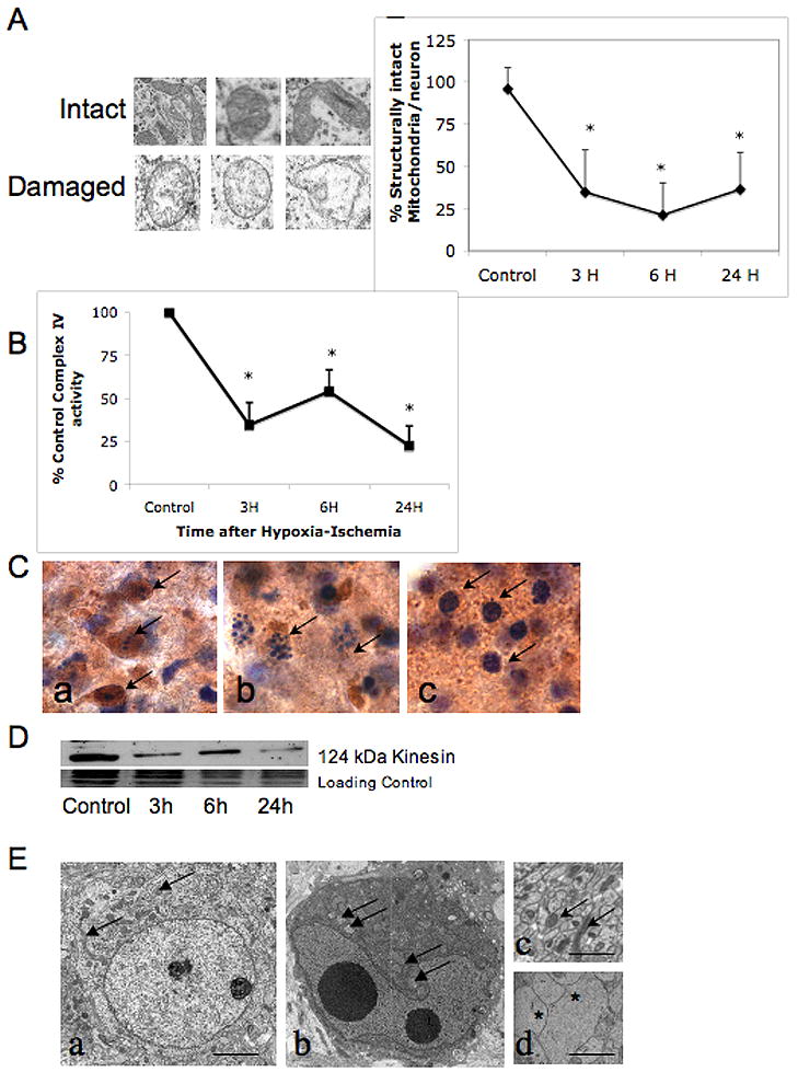
A. Individual mitochondria from striatal neurons were classified as having intact inner cristae and inner and outer membrane or swollen and disrupted inner cristae and separation of the inner and outer membrane as shown. Using these criteria, percent intact mitochondria within individual striatal neurons was calculated in uninjured control neurons and at 3, 6, and 24 hours after HI (graph, mean± SD, n =25 at each time point, * p≤ 0.05 vs control, ANOVA with Dunnett’s post hoc test). B. Mitochondrial complex IV activity rapidly declines within the first 3 hours following neonatal HI (mean ±SD of % control activity, n= 6 samples at each time point, * p≤ 0.05 vs control, ANOVA with Dunnett’s post hoc test). Partial recovery at 6 hours is not sustained and there is more profound secondary decrease at 24 hours. C. Dying striatal neurons with different cell death phenotypes have varying preservation of cytochrome oxidase histochemical activity. Cells with classic apoptotic phenotype maintain intense cytochrome c oxidase activity (Ca, brown immunoreactivity, arrows) compared to cells with incomplete chromatin condensation, consistent with ‘continuum’ phenotype (Cb) and cells with irregularly and minimally condensed chromatin (Cc).
D. Immunoblot showing loss of kinesin protein as soon as 3 hours after HI. This coincides with the loss of structural integrity and functional activity of mitochondria following neonatal HI (panels A and B). E. Healthy neurons from control striatum have intact cytoplasmic and nuclear membranes, well dispersed chromatin with nucleoli visible, intact mitochondria dispersed throughout cytoplasm (Ea, scale bar=2μm). In contrast, in injured neurons, perinuclear clustering of mitochondria occurs within darkened and condensed cytoplasm, possibly from loss of anterograde trafficking of mitochondria (Eb) (mitochondria indicated with arrows in Ea, Eb, and Ec) (Al-Abdulla and Martin, 1998, Northington et al., 2001a). Healthy appearing mitochondria are abundant in cross sections of axons of uninjured neurons (Ec) and almost completely absent in swollen injured axons (Ed, asterisks) at 6 hours following HI (scale bars =1μm).