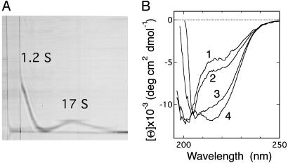
Fig. 1.
Association of the 0SS variant. (A) A Schlieren pattern, taken 42 min after the start of sedimentation. Protein concentration was 7.6 mg·ml–1 in 50 mM sodium maleate (pH 2.0). (B) Far-UV CD spectra of the 0SS solution incubated for 1 day at 25°C in 5 mM sodium maleate (pH 2.7)/45 mM sodium chloride at protein concentrations of 1, 2, 3, and 4 mg·ml–1 (labeled on each spectrum).

