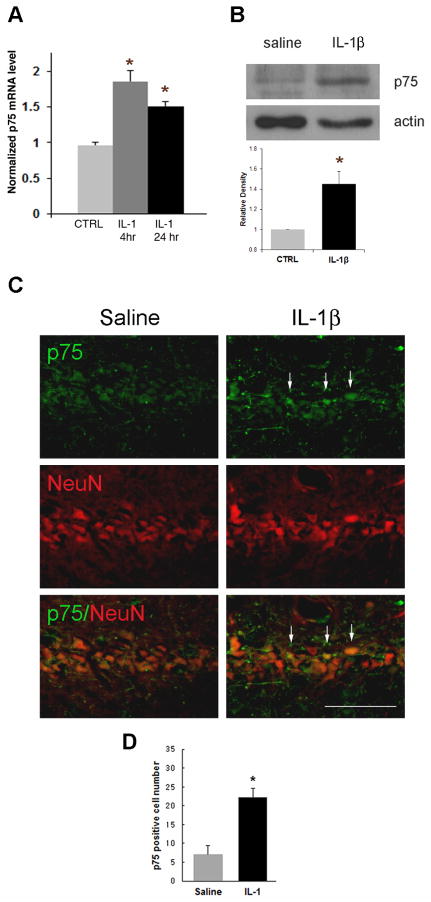
Figure 1. Unilateral IL-1β infusion increases p75NTR expression in vivo.
Rats were cannulated 7 days before infusion with IL-1β (10ng). A. p75NTR mRNA was induced by IL-1β. Tissue was taken after 4 hr or 1d treatment with IL-1β (mean±SEM, n = 3). Asterisk denotes difference from saline (p<0.05). B. 2 days after the infusion, each hippocampus was taken for Western blot assay. p75NTR expression was increased with IL-1β infusion. Quantification of blots from three different experiments and densitometric values were normalized to actin and are expressed relative to the saline control (CTRL). Error bars represent SEM. * P<0.05 relative to CTRL, two-tailed t test. C. 2 days after the IL-1β infusion, brains were perfused, sectioned through the hippocampus, immunostained with anti-p75NTR (green) and anti-NeuN (red). IL-1β infusion increased p75NTR expression (arrows) in the CA1 region of the hippocampus (right column) compared to saline infusion (left column). Scale bars = 50μm. n=6. D. Counts of p75NTR-positive cells indicated a 3-fold increase following IL-1β infusion.

