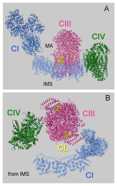
Figure 2.

Pseudo-atomic model shows the arrangement of CI, CIII and CIV in the bovine respirasome (PDB 2YBB, (Althoff et al., 2011)). (A) Side view and (B) view from IMS. Two CL molecules (in yellow) in the external cavity formed by cytochrome c1, cytochrome b and closed by chain G in each monomer of CIII (PDB 1PP9) are shown. MA denotes the mitochondrial matrix.
