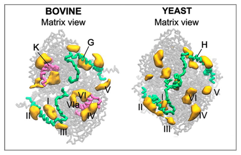
Figure 4.

CL binding sites of bovine and yeast CIII extracted from CGMD simulation of the complexes embedded in a CL/PC bilayer. CLs bound in the sites I and VI/VIa correspond to tightly bound CLs found in the crystal structures of CIII from yeast and bovine. Site I corresponds to the CL binding site in the external cavity closed by chain H in yeast and its homolog chain G in bovine. Sites VI/VIa corresponds to the conserved site for tightly bound CL in the inner cavity located close to the CIII homodimer interface (CN5, PDB 3CX5 for yeast and CL3 in PDB 1SQP for bovine). Chain K is present in the bovine CIII close to sites VI and VIa. However, there is no homolog for this chain in yeast CIII. Sites II, III, IV and V are the sites on the membrane-exposed surfaces of CIII. The figure was adapted with permission from (Arnarez et al., 2013b). Copyright (2013) American Chemical Society.
