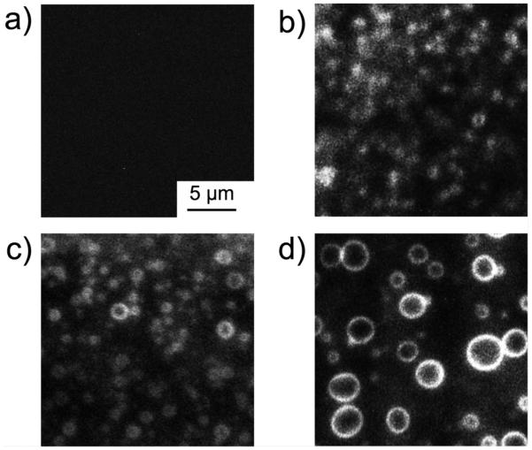
Fig. 4.
Visualization of curcumin localization at the domain boundaries. TIRF images of the planar lipid bilayer consisting of DOPC / SM / Chol were acquired (a) before the addition and at (b) 30, (c) 45 and (d) 60 minutes after the addition of curcumin. Curcumin was excited at 405 nm by a diode laser. [curcumin] / [lipid] = 8 ×10⁻⁶ at 25 °C.

