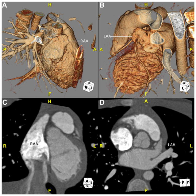
Fig. 1.
CTA images of atrial appendage anatomy in a healthy child with situs solitus. (A and B) Images are 3D volume-rendered reconstructions viewed from a right and left anterolateral position, respectively. (A) The right-sided morphologically RAA is shown, which is broad based and triangular. (B) A left-sided morphologically LAA is shown, which is narrow and finger-like. (C) This oblique coronal image shows a normal RAA. Note that the pectinate muscles extend into the body of the atrium. (D) This oblique transverse image shows a normal LAA, with narrow, smooth-walled neck. A, anterior, CTA, CT angiography; F, feet; H, head; L, left; LAA, left atrial appendage; P, posterior; R, right; RAA, right atrial appendage; 3D, 3-dimensional.

