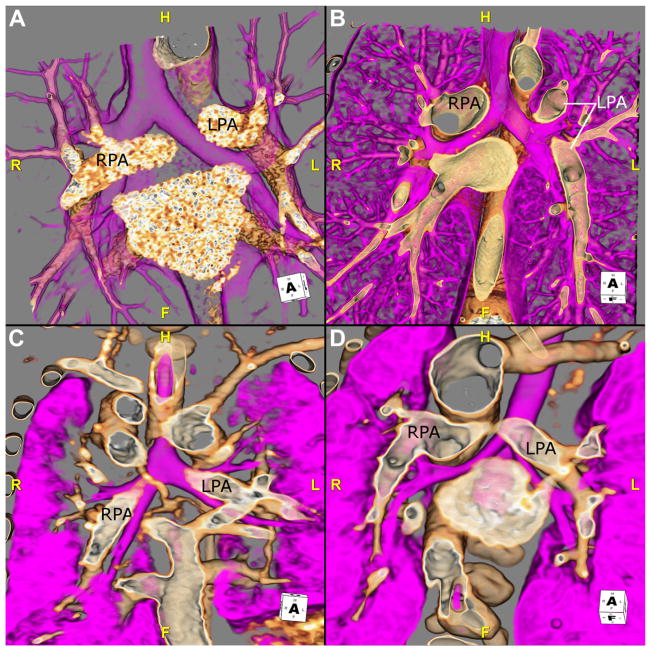
Fig. 2.
The relationship of the bronchus to its ipsilateral pulmonary artery can be useful in determining situs. These images are 3D volume-rendered CTA reconstructions in 4 different patients. Each image shows the airway (pink) and the RPA and LPA. (A) This image shows this relationship in a patient with situs solitus. The right bronchus is eparterial; the bronchus branches superior to the first lobar division of the pulmonary artery. The left bronchus is hyparterial; the bronchus branches inferior to the first lobar division of the pulmonary artery. (B) The relationship of the bronchi and the pulmonary arteries RPA and LPA of a patient with situs inversus is shown. The right-sided bronchus is hyparterial, consistent with a morphologic left bronchus, whereas the left-sided bronchus eparterial is consistent with a morphologic right bronchus. (C) This image shows a patient with right isomerism. Both bronchi are eparterial. (D) This image is from a patient with left isomerism, showing bilateral hyparterial bronchi. CTA, CT angiography; F, feet; H, head; L, left; LPA, left pulmonary artery; R, right; RPA, right pulmonary artery; 3D, 3-dimensional.

