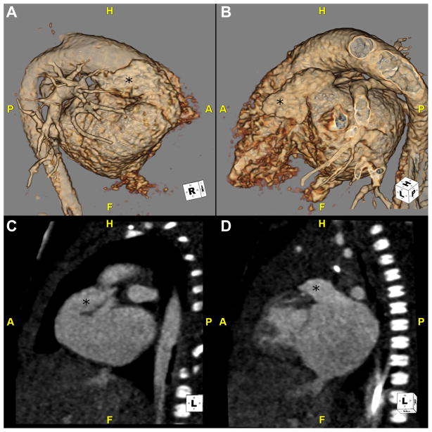
Fig. 7.
This is a CTA in a neonate with left isomerism. (A and B) These 3D volume-rendered reconstructions are viewed from a right and left anterolateral position, respectively. (C and D) These sagittal images are through the right-sided and left-sided atrial appendages, respectively. The appendages (*) look similar and show characteristics of a morphologic left atrial appendage. They are narrow, finger-like structures. A, anterior; CTA, CT angiography; F, feet; H, head; P, posterior.

