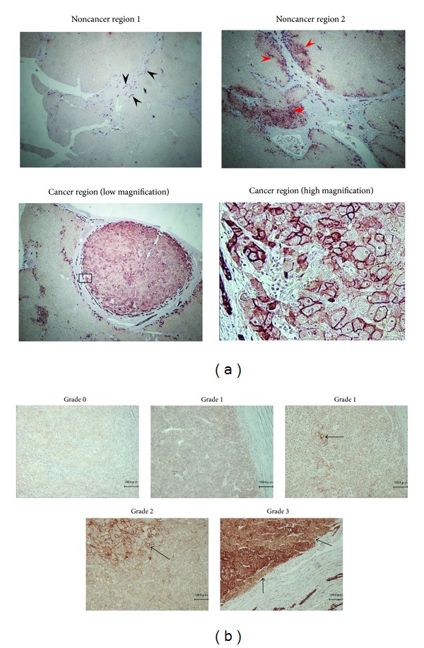
Figure 1.

Immunohistochemical staining of EpCAM in resected HCCs. (a) EpCAM expression was observed in bile duct (black arrows). Many hepatocytes did not express EpCAM, but it was expressed in the regenerated damaged liver tissue like that caused by cirrhosis (red arrows). EpCAM expression in hepatocellular carcinoma (under panels). (b) Black arrows indicate cells with high EpCAM expression in HCC. EpCAM (Grade 0 (0%), Grade 1 (<10%, or diffusely weakly expression), Grade 2 (≥10% and <50%, resp.), and Grade 3 (≥50%)).
