
Figure 3.
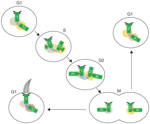
Centriole and centrosome asymmetries. Schematic representation of centrioles (green), with distal and subdistal appendages (triangles), G1–G2 tether (blue), S–M linker (red) and pericentriolar material associated with the base of each centriole. The centrioles are numbered to indicate their origin and age. The centriole marked ‘1’ (centriole 1) is the older of the two centrioles in the G1 cell at the upper left. Centriole 2 formed in the previous cell cycle, as a procentriole adjacent to centriole 1. The centrioles in this cell are disengaged (no S–M linker), but tethered (G1–G2 tether). In S phase new procentrioles grow from each of centrioles 1 and 2 and elongate in G2 phase. These new centrioles (3 and 4) are engaged to their mother centrioles (1 and 2, respectively), but are otherwise equivalent. Centriole 2 acquires appendage proteins at the G2/M transition and appendages proper in the subsequent G1. The two centrosomes segregate at mitosis, with one cell receiving the 1, 3 pair and the other receiving the 2, 4 pair. Although the centriole pairs are morphologically equivalent, there is a functional difference, in that the cell receiving the older mother centriole (centriole 1), is able to form a primary cilium earlier in the cell cycle than the other cell. The pericentriolar material at the base of each centriole is represented in different colours to indicate the possibility that proteins associated with the centrioles might be asymmetrically segregated at mitosis with them.