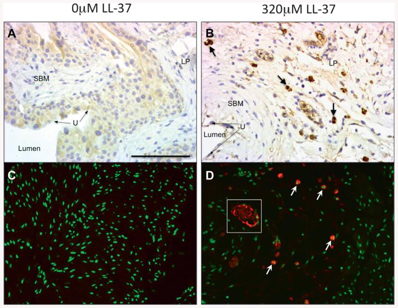
Figure 4.
Immunohistochemistry for mast cell tryptase. Brown areas represent mast cell detection (A and B). LP, lamina propria. SBM, bladder smooth muscle. U, urothelium. DAB stain, reduced from ×20. Immunofluorescence (C and D). Red areas represent mast cell detection. No evidence of mast cell presence in nonchallenged tissues (A and C). Robust mast cells in LL-37 challenged tissues (B and D). Arrows indicate mast cell samples. White square indicates representative tryptase degranulation pattern in endovasculature. Scale bar represents 75 μM (A). Reduced from ×20.

