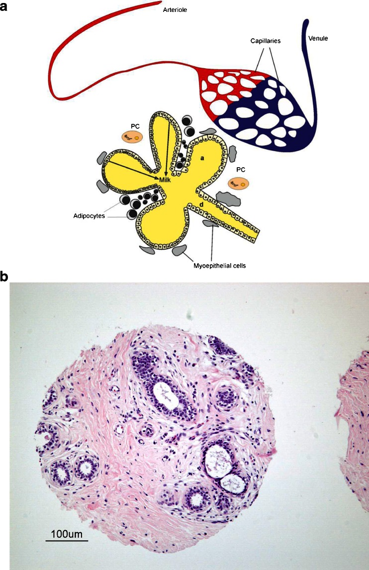
Fig. 2.
a Model alveolus (a) with subtending duct (d) showing blood supply, adipocyte stroma, myoepithelial cells, and plasma cells (PC). b Histology of human mammary gland. The sample shown in this figure is from a tissue microarray developed by the Cooperative Human Tissue Network (CHTN) of the National Cancer Institute (http://www.chtn.nci.nih.gov/). Mammary gland alveoli and ducts form an extensive network of interconnected structures

