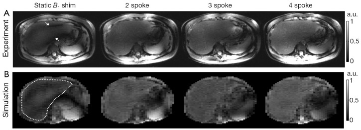
Figure 3.

Comparison of static B1 shimming and multi-spoke pTX pulse design when used to mitigate B1+ inhomogeneity in liver MRI at 7T. (A) central axial views (1 mm thick) of 3D GRE datasets acquired in one subject using four RF pulse designs. Note that two dark stripes (as indicated by white arrows) observed with the static B1 shimming solution were effectively removed by using multi-spoke pTX RF pulses; (B) corresponding Bloch simulations of the transverse magnetization using the same pulses as in (A). The white dotted loop in the leftmost image indicates the ROI covering the liver area for which B1+ homogenization was intended. Note the good agreement between experiments and simulations in terms of excitation pattern characteristics.
