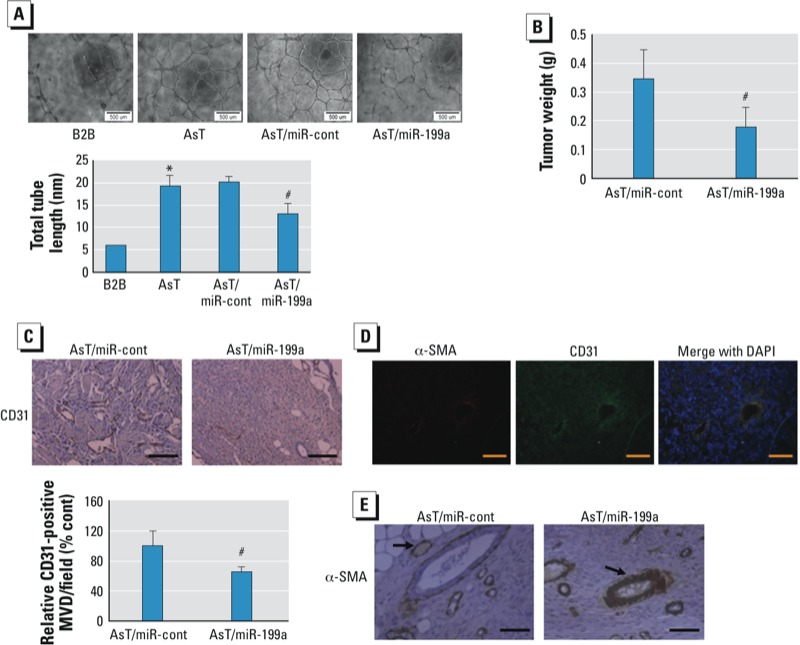
Figure 2.

miR-199a inhibits arsenic-induced angiogenesis. (A) HUVEC cells were cultured in serum-free medium overnight and resuspended in basic EBM-2 medium. Data represent mean ± SE from six replicates from each treatment and were analyzed by one-way ANOVA. Bar = 500 μM. (B) Weight of xenograft tumors from nude mice was measured 6 weeks after cell injection. Data represent mean ± SE (n = 10/group). (C) Paraffin-embedded tumor tissue sections from both groups were used for immunohistochemical staining using antibodies against CD31. Top: representative sections (magnification: 160×; bar = 50 μM). Bottom: quantification of microvessel density (MVD) indicated by CD31 staining in tumor sections. Data represent mean ± SE from five different tumor sections from each group. (D) Frozen tissue sections were used for immunofluorescence staining (magnification: 200×; bar = 50 μM). (E) Paraffin-embedded tumor tissue sections were used for immunohistochemical staining using antibodies against α-SMA. Representative sections are shown. Arrow indicates mural cells (magnification: 320×; bar = 50 μM). *p < 0.05, compared with B2B. #p < 0.05, compared with AsT/miR-cont.
