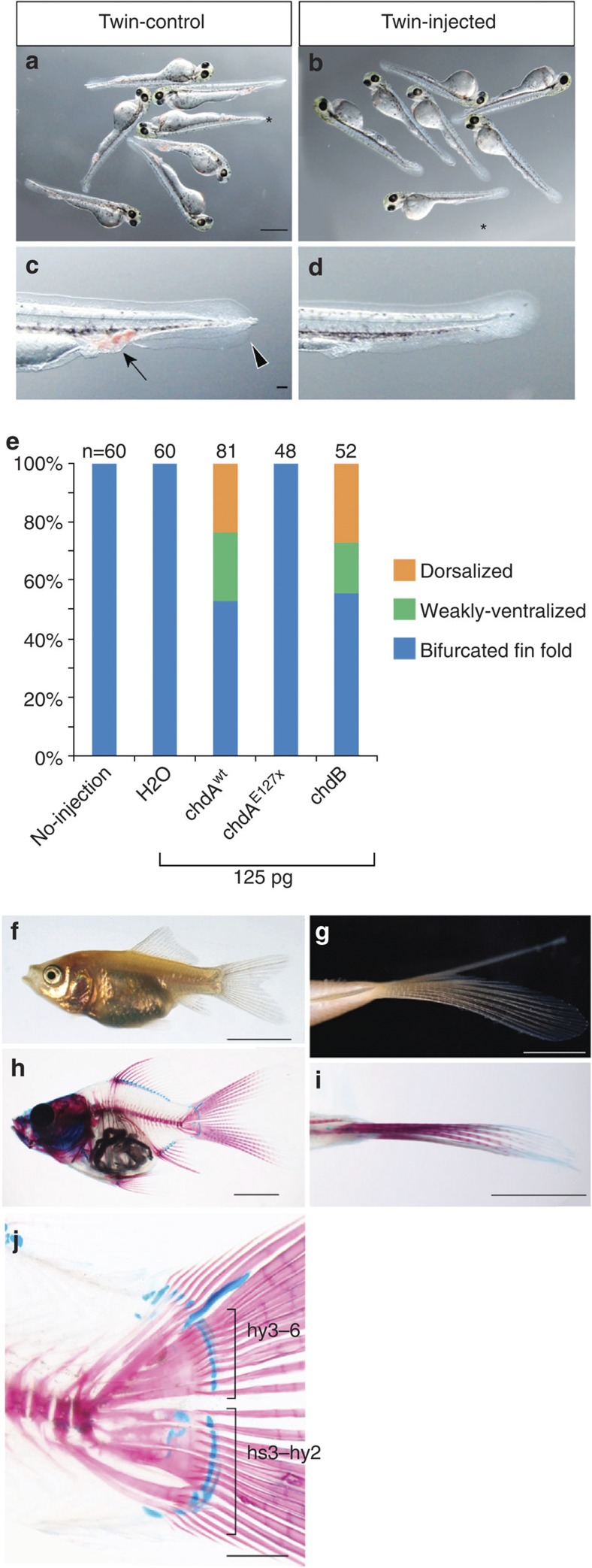
Figure 3. Rescue of the twin-tail phenotype by mRNA microinjection.
(a,b) Larval phenotypes of twin-tail goldfish. Non-injected controls (a) or embryos injected with 125 pg chdAwt mRNA (b). (c,d) Magnified view of the caudal regions indicated by asterisks in a and b, respectively. Arrow and arrowhead indicate accumulated blood cells and bifurcated fin folds, respectively. (e) Proportion of rescued specimens following injection of embryos with the indicated mRNA. The number of larvae analysed is indicated above each bar. (f–j) Twin-tail goldfish juvenile injected with chdAwt mRNA. (f) Lateral view. (g) Ventral view of the caudal level. (h) Lateral view of the skeletal structure. (j) Magnified view of the caudal region of h. The dorsal phenotype criteria were based on previous descriptions. Panels a and b, and c and d are at the same magnification, respectively. Scale bars, 1 mm (a,h,j), 100 μm (c), 5 mm (f–i).

