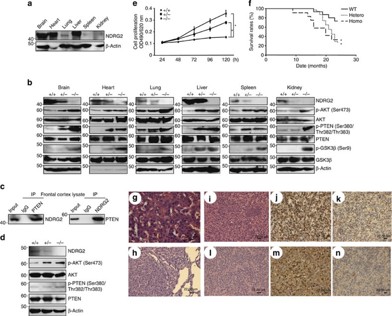
Figure 6. NDRG2-deficient mice are susceptible to tumour formation.
(a) Western blots of 3-month-old adult tissues of WT mice. The data are representative of two experiments. (b) Western blots of various tissues of 3-month-old NDRG2-deficient mice. The data are representative of two experiments. (c) The co-immunoprecipitation of PTEN and NDRG2 in frontal cortex homogenates from 3-month-old WT mice. The data are representative of three experiments. (d) Whole-cell lysates from WT, NDRG2+/− and NDRG2−/− embryonic fibroblasts were subjected to western blotting. (e) The proliferation rates of WT, NDRG2+/−, and NDRG2−/− embryonic fibroblasts. The mean±s.d. is shown; *P<0.05 (Student’s t-test). The data are representative of three experiments. (f) Kaplan–Meier survival curves of WT (n=15), NDRG2+/− (n=31) and NDRG2−/− (n=12) mice up to 24 months of age. The difference in survival was statistically significant between WT and NDRG2+/− or NDRG2−/− mice (*P<0.05, log-rank test). (g) A hepatocellular carcinoma section from an NDRG2+/− mouse liver was subjected to H&E staining. The results indicated the presence of a large sheet of hepatic cords composed of several hepatocytes of variable nuclei and cell sizes; the nuclei of the carcinoma cells were hyperchromatic with prominent nucleoli (scale bar, 22 μm). (h) A bronchoalveolar carcinoma section from an NDRG2+/− mouse lung was subjected to H&E staining. The histopathology demonstrated a well-circumscribed mass of a solid sheet of neoplastic cells containing hyperchromatic nuclei with signs of frequent mitosis and an indistinct basophilic cytoplasm (scale bar, 22 μm). (i–k) Lymphoma sections from the mesenteric lymph node of an NDRG2+/− mouse. The histopathology indicated the presence of diffuse pleomorphic large lymphoid cells with vesicular nuclei, prominent nucleoli and scant cytoplasm (i). Robust staining for CD3 was present in the cytoplasm (j), but B220 staining was negative (k) (scale bar, 22 μm). (l–n) Lymphoma sections from an NDRG2+/− mouse spleen were examined by histopathology. The results demonstrated pleomorphic large lymphoid cells with vesicular nuclei, prominent nucleoli, some nuclear distortion and numerous cells undergoing mitosis (l). Staining for CD3 revealed robust cytoplasmic staining in cells, ranging from white pulp to red pulp (m). B220 staining was negative (n) (scale bar, 22 μm).

